Product
Dyes
Reverse staining of SDS-PAGE in only 5 minutes — no destaining or heating required.
VisPRO™ utilizes an innovative imidazole–zinc ion precipitation reaction within the gel matrix to visualize protein bands rapidly and clearly. The process is complete in just 5 minutes, delivering results comparable to silver or SYPRO Ruby staining while avoiding the tedious steps of traditional methods.
Special Characteristic :
Ultra-Fast Protocol – Complete protein visualization within 5 minutes, ready for imaging or excision.
High Sensitivity – Detects as low as 1 ng of protein, matching silver stain and SYPRO Ruby performance.
Protein-Safe Mechanism – Stains the gel matrix, not the protein, ensuring no interference with protein integrity or downstream analysis.
Downstream Compatibility – The stained bands can be directly excised and used for Western blotting, LC-MS/MS, enzyme digestion, and other analytical workflows.
Environmentally Friendly – The staining solution is non-toxic, non-biohazardous, and safe for both users and the environment.
Long-Term Stability – Highly stable formulation with consistent results and low background over extended use.
Order Information :
| Cat. No. | Product Name | Description |
| VP01-125 | VisPRO™ 5 Minute Protein Stain Kit |
1 kit |
| VP01-500 | VisPRO™ 5 Minute Protein Stain Kit |
1 kit 500ml solution 1 + 500ml solution 2 |
| VP05-125 | VisPRO™ 5 Minute Protein Stain Kit |
1 kit 5X concentrate solution 125ml solution 1 + 125ml solution 2 |
| VP05-500 | VisPRO™ 5 Minute Protein Stain Kit |
1 kit 5X concentrate solution 500ml solution 1 + 500ml solution 2 |
Product Information :
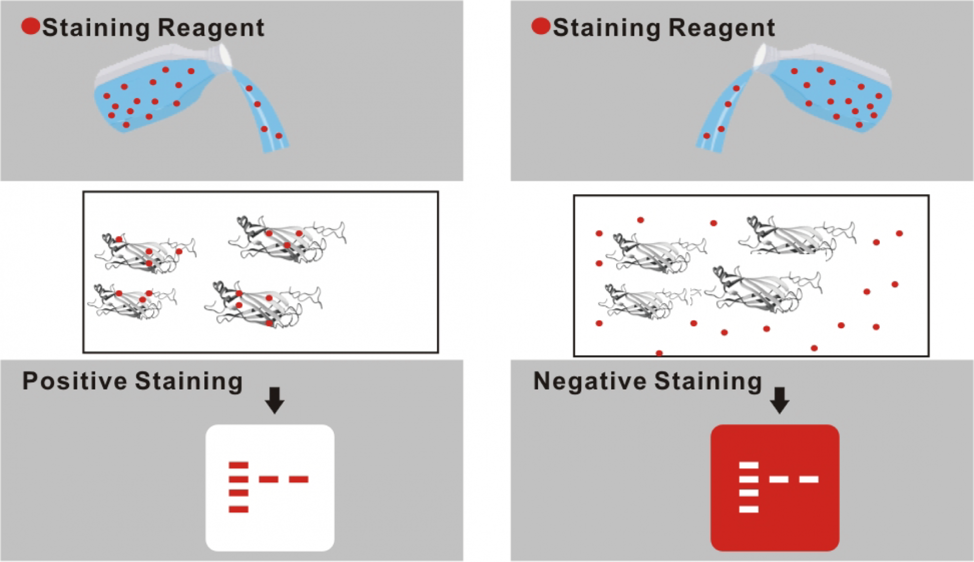
Figure 1: Positive staining vs Reverse staining.
In positive staining methods such as Coomassie or silver staining, the protein bands absorb dye, appearing as dark bands on a clear background. The staining reagent interacts directly with proteins, which can sometimes alter protein structure or interfere with downstream analysis.
In contrast, reverse staining (as used in VisPRO™ 5 Minutes Protein Stain Kit) relies on imidazole–zinc ion precipitation within the gel matrix. Here, the gel background becomes opaque white, while protein bands remain clear and sharply defined.

Figure 2: Operation workflow
The VisPRO™ workflow is designed for simplicity and speed. After electrophoresis, gels are immersed directly in the ready-to-use staining solution in a black background container—no fixation, heating, or destaining required. Within five minutes, protein bands or spots become clearly visible at the nanogram sensitivity level.
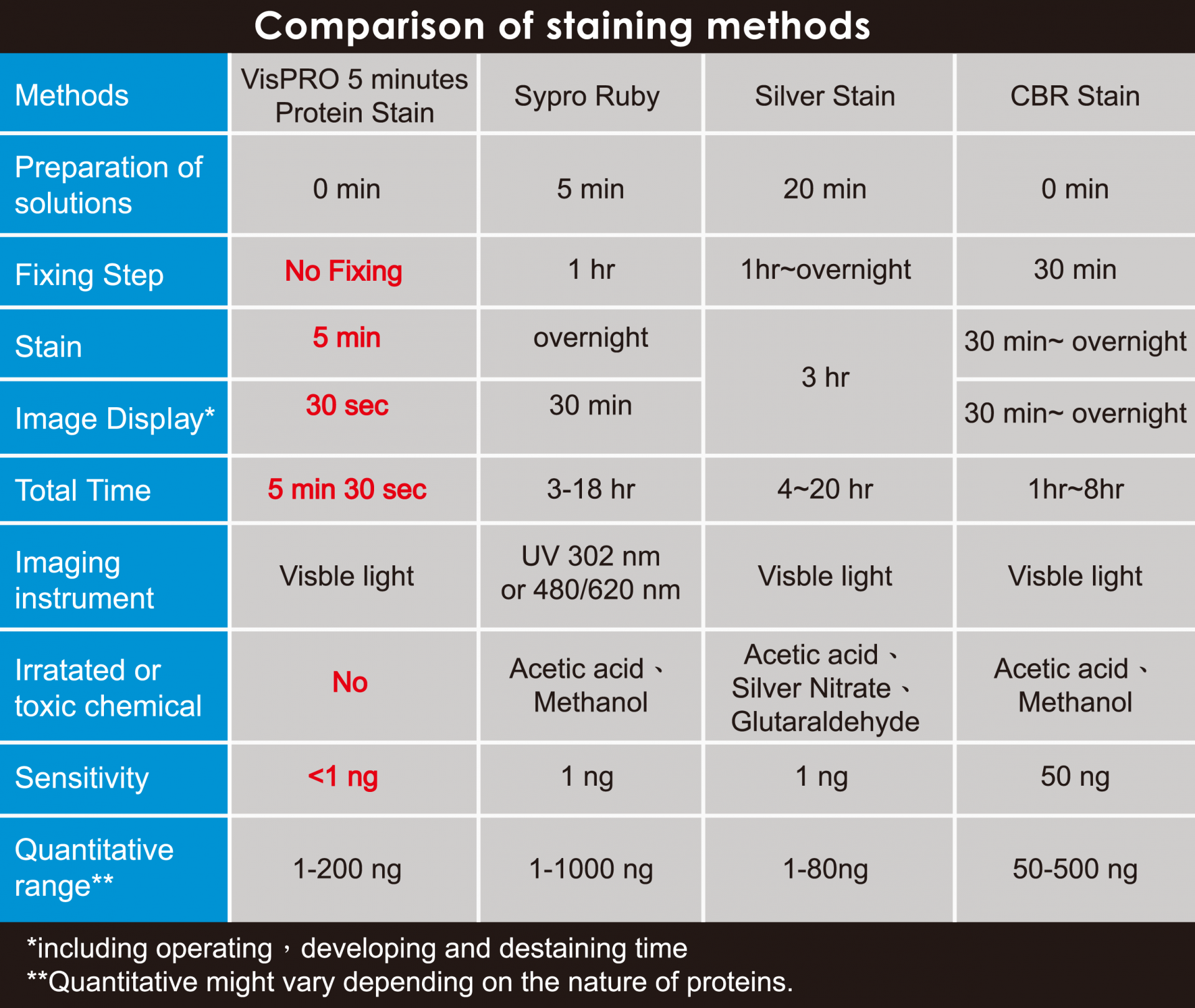
Figure 3: Comparison of staining methods
VisPRO™ produces sharp, high-contrast bands comparable to silver and SYPRO Ruby staining — but in a fraction of the time and with full compatibility for downstream analysis, making it an ideal choice for fast, safe, and quantitative protein visualization.

Figure 4: SDS-PAGE Gel Stained with VisPRO™ 5 Minutes Protein Stain Kit
The image shows an SDS-PAGE gel stained with the VisPRO™ 5 Minutes Protein Stain Kit, captured using a standard cellphone camera without any image enhancement. Even under non-laboratory imaging conditions, the clear, high-contrast protein bands are easily visible, demonstrating the strong staining performance and uniform background of the VisPRO™ reverse-staining system.
Reference:
| 1 | Liau, Y. J., Wen, L., Shaw, J. F. & Lin, C. T. A highly stable cambialistic-superoxide dismutase from Antrodia camphorata: expression in yeast and enzyme properties. J Biotechnol 131, 84-91 (2007). |
| 2 | Chen, S. C. et al. Acute hypoxia enhances proteins' S-nitrosylation in endothelial cells. Biochem Biophys Res Commun 377, 1274-1278 (2008). |
| 3 | Kizuka, Y. et al. Laminin-1 is a novel carrier glycoprotein for the nonsulfated HNK-1 epitope in mouse kidney. Glycobiology 18, 331-338 (2008). |
| 4 | Wu, S. Y., Chin, L. T., Chen, L. M. & Chen, H. M. Direct visualization of fluorescent signals in protein gels using a backlit blue light plate. Proteomics 8, 3382-3388 (2008). |
| 5 | Huang, B., Chen, S. C. & Wang, D. L. Shear flow increases S-nitrosylation of proteins in endothelial cells. Cardiovasc Res 83, 536-546 (2009). |
| 6 | Huang, B., Liao, C. L., Lin, Y. P., Chen, S. C. & Wang, D. L. S-nitrosoproteome in endothelial cells revealed by a modified biotin switch approach coupled with Western blot-based two-dimensional gel electrophoresis. J Proteome Res 8, 4835-4843 (2009). |
| 7 | Lin, C. Y. et al. A comprehensive evaluation of imidazole-zinc reverse stain for current proteomic researches. Proteomics 9, 696-709 (2009). |
| 8 | Wu, H. C. et al. Isoelectric focusing management: an investigation for salt interference and an algorithm for optimization. J Proteome Res 9, 5542-5556 (2010). |
| 9 | Wu, H. C., Yen, C. C., Tsui, W. H. & Chen, H. M. A red line not to cross: evaluating the limitation and properness of gel image tuning procedures. Anal Biochem 396, 42-50 (2010). |
| 10 | Chen, Yun-An, et al. "Mercury-induced biochemical and proteomic changes in rice roots." Plant Physiology and Biochemistry 55 (2012): 23-32. |
| 11 | Chiu, Cheng‐Di, et al. "Investigation of the effect of hyperglycemia on intracerebral hemorrhage by proteomic approaches." Proteomics 12.1 (2012): 113-123. |
| 12 | Huang, Bin, et al. "The role of nitric oxide on rosuvastatin-mediated S-nitrosylation and translational proteomes in human umbilical vein endothelial cells." Proteome science 10.1 (2012): 43. |
| 13 | Chen, Lei‐Chin, et al. "Molecular mechanisms of 3, 3′‐dichlorobenzidine‐mediated toxicity in HepG2 cells." Environmental and molecular mutagenesis (2014). |
| 14 | Huang, Bin, et al. "Arsenic Modulates Posttranslational S-Nitrosylation and Translational Proteome in Keratinocytes." The Scientific World Journal (2014). |
| 15 | Lin, Ming Chung, et al. "Rosuvastatin Modulates the Post-Translational Acetylome in Endothelial Cells." Acta Cardiologica Sinica 30.1 (2014): 67-73. |
| 16 | Yih-Huei Uen, et al. "Analysis of differentially expressed novel post-translational modifications of plasma apolipoprotein E in Taiwanese females with breast cancer. " Journal of Proteomics (2015) 126, 3: 252-262. |
| 17 | Yifan Hu, et al. “Cellular splicing factor UAP56 stimulates trimeric NP formation for assembly of functional influenza viral ribonucleoprotein complexes” Scientific Report (2017) 7: 14053. |
| 18 | Chen H-M, et al. “The application of post-translational modification oriented serum proteomics to assess experimental diabetes with complications.” PLoS ONE (2018) 13, 11: e0206509. https://doi.org/10.1371/journal.pone.0206509 |
| 19 | Cheng-Ju Kuo, et al. “A multi-omic analysis reveals the role of fumarate in regulating the virulence of enterohemorrhagic Escherichia coli” Cell Death & Disease (2018) 9: 381 |
| 20 | Chiao-Yin Sun, et al. “A novel SNP in the 5’ regulatory region of organic anion transporter 1 is associated with chronic kidney disease.” Scientific Reports (2018) 8: 8085 |
| 21 | Heng-Dao Lin, et al. “Proteomic analysis of ametryn toxicity in zebrafish embryos.” Environmental Toxicology (2018) 33, 5 |
| 22 | Shao-hsuan Wen, et al. “Sulbactam-enhanced cytotoxicity of doxorubicin in breast cancer cells” Cancer Cell International (2018) 18:128 |
| 23 | Liao, Chao-Tsai, Ying-Chuan Chiang, and Yi-Min Hsiao. "Functional characterization and proteomic analysis of lolA in Xanthomonas campestri s pv. campestris." BMC microbiology 19.1 (2019): 1-18. |
| 24 | Lo, Hsueh-Hsia, et al. "The clpX gene plays an important role in bacterial attachment, stress tolerance, and virulence in Xanthomonas campestri s pv. campestris." Archives of Microbiology 202.3 (2020): 597-607. |
| 25 | Ko, Yuan-Tih, et al. "White muscle proteome analysis showing insights into the protein expression in orange-spotted grouper (Epinephelus coioides) muscle." Food Bioscience (2020): 100655. |
| 26 | Lin, Tung-Yi, et al. "Functional proteomic analysis reveals that fungal immunomodulatory protein reduced expressions of heat shock proteins correlates to apoptosis in lung cancer cells." Phytomedicine 80 (2021): 153384. |
| 27 | Huang, Yu-Jen, et al. "Umbilical cord blood plasma-derived exosomes as a novel therapy to reverse liver fibrosis." Stem Cell Research & Therapy 12.1 (2021): 1-13. |
| 28 | Lin, Chia-Hung, et al. "Comparative O-GlcNAc Proteomic Analysis Reveals a Role of O-GlcNAcylated SAM68 in Lung Cancer Aggressiveness." Cancers 14.1 (2022): 243. |
| 29 | Lin, You-Cian, et al. "Differential Serum Proteomic Signatures between Acute Aortic Dissection and Acute Myocardial Infarction." Biomedicines 11.1 (2023): 161. |
| 30 | Liao, Chien-Sen, et al. "Impacts of endocrine disruptor di-n-butyl phthalate ester on microalga Chlorella vulgaris verified by approaches of proteomics and gene ontology." Molecules 25.18 (2020): 4304. |